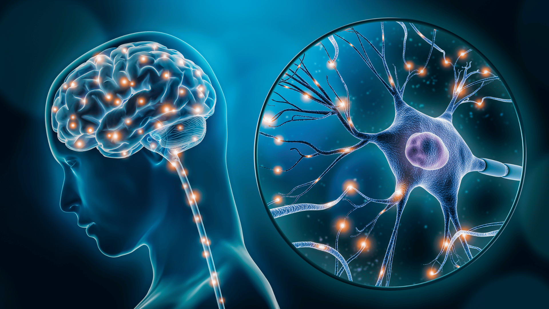
Johns Hopkins University researchers developed a breakthrough organoid that models the whole brain with neural tissues and rudimentary blood vessels. The brain organoids could change how scientists study disorders like autism and schizophrenia.
“Most brain organoids that you see in papers are one brain region, like the cortex or the hindbrain or midbrain,” said the study’s lead author, Annie Kathuria. “We’ve grown a rudimentary whole-brain organoid; we call it the multi-region brain organoid (MRBO).”
Multi-Region Brain Organoid

Organoids are mini, simplified versions of organs created in a lab. Scientists grow 3D tissues to mimic functioning organoids. Because the cells are derived from a patient, they help doctors and scientists understand a disease or tailor treatments.
Researchers from Johns Hopkins University say this is one of the first times that scientists have been able to generate an organoid with tissues from different brain regions to develop a coordinated network.
Advertisement
Kathuria and her team created this complex model using neural cells from different brain regions and rudimentary blood vessels in separate lab dishes. They used sticky proteins to join individual parts together, which allowed the tissues to form connections and begin electrical activity as a network.
As a result, researchers say that the mini brain has a cell-type range similar to that of a 40-day-old human fetus, with about 80% of the normal cell types expressed.
According to the research team, the multi-region, lab-grown brain offers a crucial platform for studying the entire brain. Kathuria said, “Whole-brain organoids let us watch disorders develop in real time, see if treatments work, and even tailor therapies to individual patients.”
Additionally, researchers say they could use the organoids to test experimental drugs and improve the low success rates of clinical trials for neuropsychiatric drugs.
“If you can understand what goes wrong early in development, we may be able to find new targets for drug screening,” Kuthuria concluded.