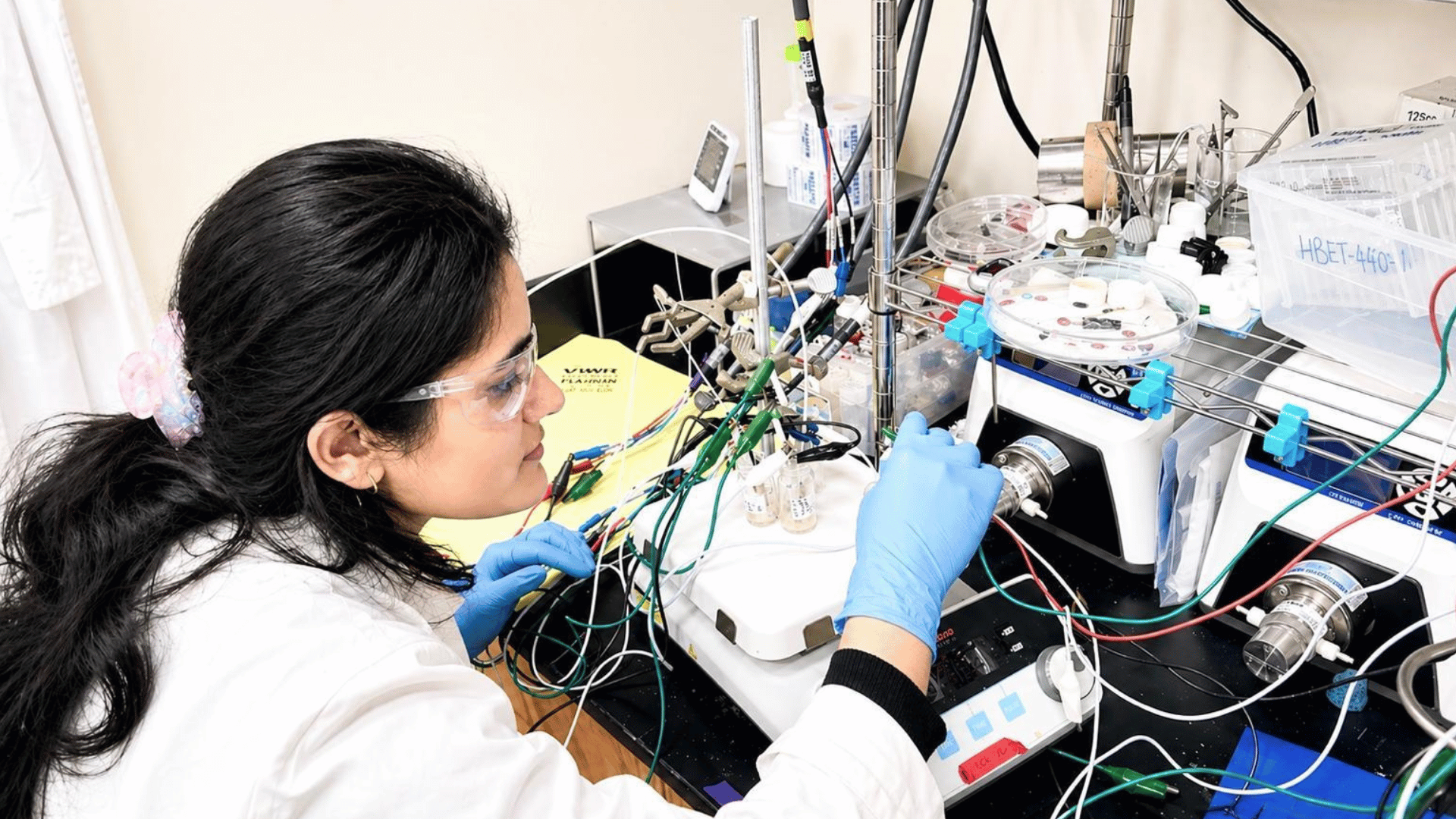
Scientists at MIT recently discovered something they didn’t think was possible. While studying high-power lasers and optical fibers, they found that light can organize itself into a perfect, needle-sharp beam. This discovery might change how we study the brain and test new drugs for diseases like Alzheimer’s or ALS.
The project started when the team pushed a standard optical fiber to its limit. Usually, when you pump a lot of power into a laser fiber, the light becomes a messy, scattered disaster. However, graduate student Honghao Cao noticed that right before the fiber was about to burn up, the chaotic light suddenly collapsed into a single, highly focused “pencil beam.”
Getting the Laser Exactly Right

To make this happen, the team had to get two things exactly right. They had to aim the laser at a perfect zero-degree angle and crank the power up until the light started interacting with the glass of the fiber itself.
“At this critical power, the nonlinearity can counter the intrinsic disorder, creating a balance that transforms the input beam into a self-organized pencil beam,” Cao explained.
Usually, getting a beam this steady and sharp requires expensive equipment and a lot of engineering. This new method, however, is much simpler. Sixian You, the senior author of the study, notes that the simplicity is the best part. “That is the charm of this method — you could do this with a normal, optical setup and without much domain expertise,” she said.
Advertisement
Better Medicine For the Brain
The researchers put their new beam to work by looking at the human blood-brain barrier. This is the layer of cells that keeps toxins out of your brain, but it also makes it hard for medicine to get in. Most imaging tools are slow, only capturing one small slice of the barrier at a time.
Using the self-organized beam, the team was able to take 3D images 25 times faster than the current gold-standard method. They could actually watch individual cells absorbing drugs in real-time without needing to use any special glowing tags on the cells.
“The common belief in the field is that if you crank up the power in this type of laser, the light will inevitably become chaotic. But we proved that this is not the case. We followed the evidence, embraced the uncertainty, and found a way to let the light organize itself into a novel solution for bioimaging,” said You.
Roger Kamm, a professor at MIT, points out that this is a big deal for the pharmaceutical industry. “For the first time, we can now visualize the time-dependent entry of drugs into the brain and even identify the rate at which specific cell types internalize the drug,” he added.
Now, the team plans to see if they can use this beam to look at neurons and eventually turn this discovery into a tool that doctors and scientists everywhere can use.