Advertisement
Scientists are in the process of finding out more about the brain and its tendencies. specifically when brain cells shut off. They’re looking at the brain’s “off switches.” This new technology tracks when brain cells switch off, usually after a burst of activity, known as inhibition. Until now, it’s been a mystery how long neurons stay active and when they turn off.
Scientists at Scripps Research came up with a new technique to study why the brain’s “off switches” go out of control in normal behavior, in diseases and disorders.
Inhibition
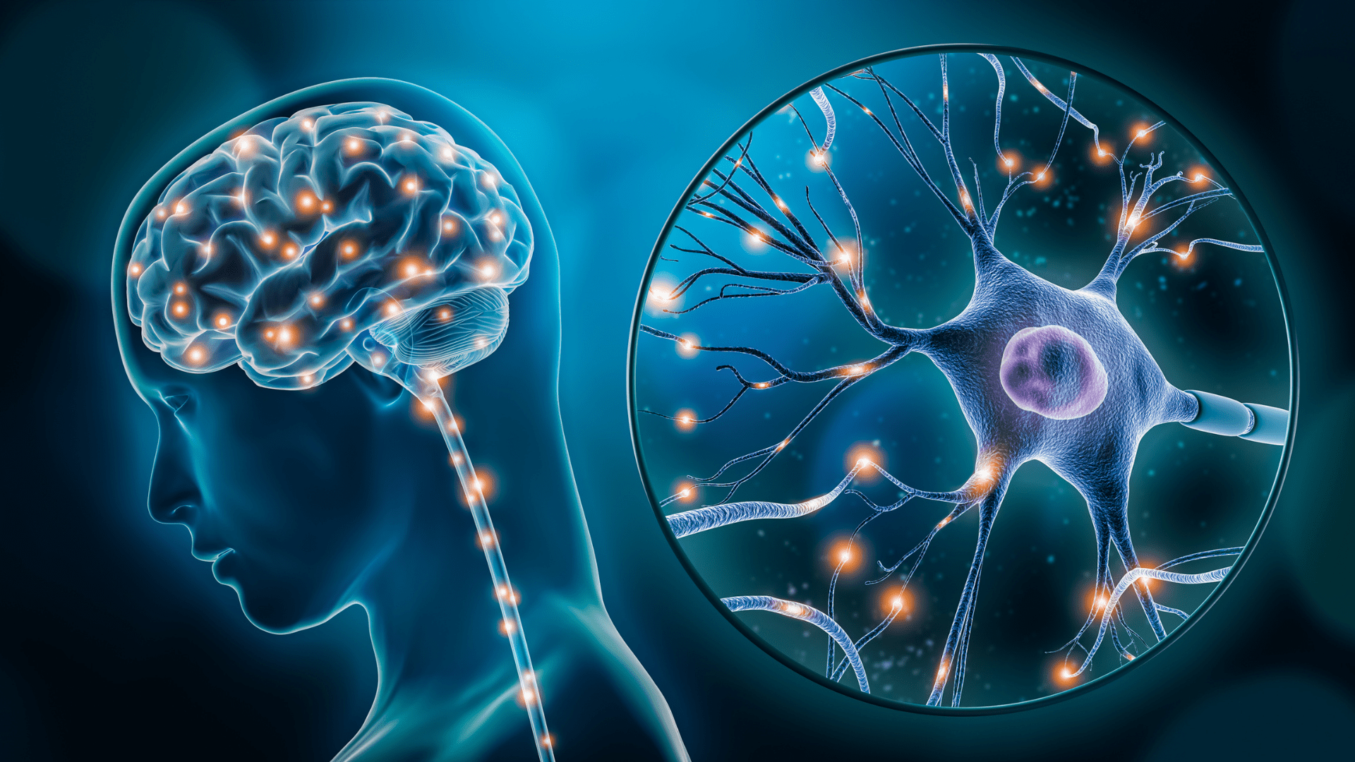
It is agreed that inhibition of neurons is how the brain regulates activities but finding a way to track it proved to be difficult. Until now, only a few have been able to look at it in a trackable way. Li Ye and John Yates are both professors at Scripps Research. Together, they are studying how brain cells change when they’re actively firing and comparing them to when they are done firing. When a brain cell fires, it is passing a message through an electrical charge.
Proteins are important in this process. To measure the levels and characteristics of the modifications of these proteins, scientists use optogenetics. This allows them to encode information from selected cells. Through this process, they identified a protein called pyruvate dehydrogenase (PDH). PDH rapidly changes immediately after brain cells are inhibited.
PDH Protein